02241282889 Helpline
 Book Appointment
What is a PET-CT Scan…

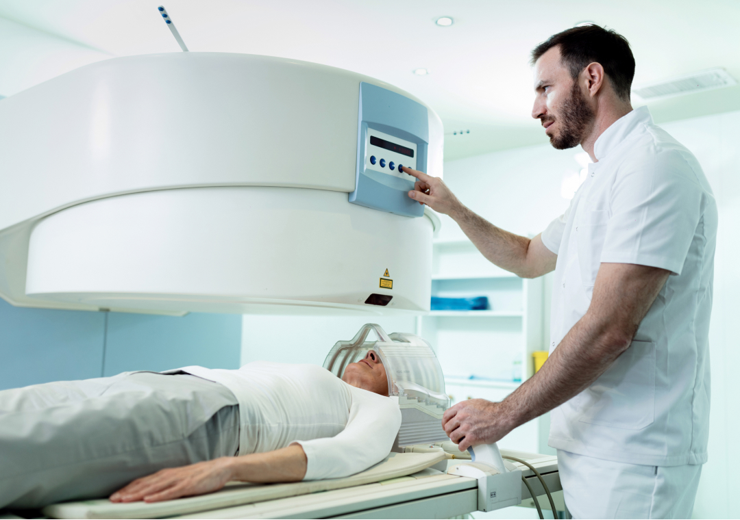
A PET CT scan for cancer is an advanced imaging test that combines functional and anatomical information to help doctors understand what is happening inside the body. It can help identify areas of increased metabolic activity, locate abnormal tissues, assess whether cancer has spread, and, when compared to previous scans, evaluate how a tumour responds to treatment.
Unlike conventional imaging alone, PET CT combines Positron Emission Tomography (PET) with Computed Tomography (CT). The PET component provides information about tissue activity, while the CT component provides detailed anatomical images. Together, these images can give doctors a more comprehensive view of cancer.
For many patients, an FDG PET CT scan is used because cancer cells may consume glucose at a higher rate than many normal tissues. A radioactive glucose-like tracer called fluorodeoxyglucose (FDG) can highlight areas of increased glucose metabolism on the scan.
However, FDG PET CT is not appropriate for every type of cancer or every patient. Doctors decide whether it is useful based on the type of cancer, suspected or known cancer, clinical history, symptoms, previous investigations, and the purpose of the examination.
A PET CT scan is a nuclear medicine imaging procedure that combines two types of imaging in a single examination.
PET shows areas of increased or decreased metabolic activity, depending on the tracer used. CT provides detailed structural information about organs and tissues.
During an FDG PET CT scan, a small amount of radioactive FDG is administered, usually through an intravenous injection. After an uptake period, the patient undergoes scanning. The PET images show tracer distribution, while CT images help identify the precise anatomical location of areas showing abnormal tracer uptake.
This combination can be particularly useful in cancer imaging because it provides both functional and anatomical information.
The role of PET CT in cancer treatment extends beyond simply looking for a tumour. Depending on the cancer type and clinical situation, doctors may use PET CT to help with:
Importantly, increased FDG uptake does not always mean cancer. Infection, inflammation and some other non-cancerous conditions can also show increased metabolic activity. Therefore, PET CT findings need to be interpreted by an appropriately qualified specialist along with the patient's clinical history and other investigations.
One of the important applications of a PET CT scan for cancer diagnosis is identifying areas of abnormal metabolic activity.
In an FDG PET CT scan, tissues that take up more FDG may appear as areas of increased activity. When interpreted alongside the CT images, doctors can determine where these areas are located and assess whether they correspond to a structural abnormality.
This information may help doctors decide whether additional investigations, such as targeted imaging or biopsy, are required.
Cancer staging describes the range of cancer in the body. Knowing the stage is important because treatment strategies can vary significantly depending on whether cancer is localized or has spread.
PET CT for cancer staging can help identify metabolically active disease in the primary tumour, nearby lymph nodes and distant sites.
For example, PET CT may help detect disease that is not clearly visible on anatomical imaging alone. This can provide additional information that may influence the overall staging assessment.
However, the usefulness of PET CT for staging varies depending on the cancer type. It is not equally sensitive or appropriate for every malignancy.
Cancer can sometimes spread from its original location to nearby lymph nodes or distant organs. This process is known as metastasis.
A PET CT scan can survey multiple areas of the body during one examination and may help identify sites that require further evaluation.
Detecting possible metastatic disease before treatment can be important because it may change the treatment strategy. For example, a patient initially considered for localized treatment may require a different approach if distant disease is identified.
The information obtained from PET CT can contribute to multidisciplinary cancer management.
Depending on the cancer type, PET CT findings may help doctors determine whether a patient may be considered for:
PET CT does not independently determine a patient's treatment. Instead, it provides imaging information that doctors consider alongside pathology, clinical findings, laboratory tests and other imaging studies.
In some situations, cancer can occur in multiple areas or there may be uncertainty about which lesion should be sampled.
PET CT may help identify a metabolically active and clinically relevant lesion. This information can sometimes assist doctors in selecting an appropriate area for biopsy.
The decision about biopsy remains with the treating medical team and depends on the patient's individual clinical circumstances.
A PET CT scan for cancer patients may be performed before treatment when doctors need to understand the extent of disease.
A baseline scan can provide information about the location and metabolic activity of known or suspected disease before treatment begins.
This baseline may also become useful later when doctors assess whether the disease has changed following treatment.
The exact timing of PET CT depends on the type of cancer, treatment plan and clinical indication.
PET CT may also be used after or during treatment in selected circumstances.
The purpose can include assessing whether previously identified disease has responded to treatment or investigating whether active disease may still be present.
For example, after chemotherapy, radiation therapy or other treatments, anatomical abnormalities can sometimes remain even when the amount of active cancer has decreased. PET CT can provide metabolic information that may help doctors interpret these findings.
However, PET CT should generally be performed at an appropriate interval after certain treatments because treatment-related inflammation can affect tracer uptake and potentially complicate interpretation.
The treating oncologist and nuclear medicine specialist determine the appropriate timing based on the clinical situation.
One of the important applications of PET CT is evaluating treatment response.
Before treatment, a tumour or other area of disease may demonstrate increased metabolic activity. Following effective treatment, metabolic activity may decrease.
Doctors can compare follow-up PET CT findings with previous examinations to assess changes in:
A reduction in metabolic activity can indicate a favourable treatment response in appropriate clinical settings. Conversely, persistent or increasing abnormal uptake may require further evaluation.
PET CT results should always be interpreted in context because inflammation and other non-cancerous processes can also cause increased FDG uptake.
The usefulness of PET CT depends on the cancer type, stage and clinical question. FDG PET CT is commonly used in the evaluation of several malignancies, some of the indications are:
PET CT may also be used for other cancers when clinically indicated.
Different cancers may require different radiotracers. For example, PSMA PET is used in appropriate patients with prostate cancer, while other specialized PET tracers may be used for specific clinical indications.
Therefore, the most appropriate PET scan should be selected by the treating physician based on the suspected or confirmed disease.
Both PET CT and conventional CT can play important roles in cancer evaluation, but they provide different types of information.
| Feature | PET CT | CT Scan |
|---|---|---|
| Main information | Metabolic + anatomical | Anatomical |
| Shows tissue activity | Yes, depending on tracer | No |
| Provides detailed anatomy | Yes | Yes |
| Cancer staging | Useful in selected cancers | Useful in many cancers |
| Treatment response | Can assess metabolic response | Can assess structural changes |
| Whole-body assessment | Possible depending on protocol | Possible depending on protocol |
The choice depends on the availability, cancer type, clinical question, and information already available from previous investigations.
Preparation requirements can vary depending on the type of PET CT examination and the tracer being used.
For an FDG PET CT scan, patients are commonly advised to fast for a specified period before the examination. Plain water may generally be allowed, but patients should follow the specific instructions provided by their imaging centre.
Patients should inform the medical team about:
For diabetic patients, preparation may require special instructions to manage blood glucose levels safely before the scan.
Patients should follow the centre's instructions carefully rather than changing medications or insulin doses on their own.
The typical PET CT scan procedure involves several steps:
The imaging team reviews the patient's medical history, referral and relevant clinical information.
A small amount of the appropriate radioactive tracer is administered, commonly through an intravenous injection.
The patient rests quietly for a period while the tracer distributes through the body. For FDG PET CT, Physical activity should generally be minimized during this period because it can influence tracer distribution.
The patient lies on the scanning table while the PET and CT images are acquired. The duration can vary depending on the protocol and the area being examined.
A qualified nuclear medicine physician reviews the images and prepares a report for the referring doctor.
A PET CT scan for cancer can provide valuable information about both the structure and metabolic activity of tissues. In selected cancers, it can support cancer staging, identify possible spread, establish a baseline before treatment, and help evaluate treatment response.
The role of PET CT in cancer treatment is therefore not limited to detecting abnormalities. It can provide information that helps the multidisciplinary medical team understand the extent and behaviour of disease and make more informed treatment decisions.
However, PET CT is not a standalone test for every cancer. The choice of scan and tracer depends on the patient's cancer type, clinical history and the specific question the treating doctor needs to answer.
If your doctor has recommended a PET CT scan, discuss the purpose of the examination, the required preparation, and the type of tracer being used with your healthcare team.
What is a PET-CT Scan…

Difference Between Pet Scan and…

Role of PSMA PET Scan…