doctor-breadcrum-nueclear

For the Doctors.

Our expertise over the years enables us to deliver cost-effective high-quality services.

Contact Us
for-doctor-the-brand-nueclear

The Brand

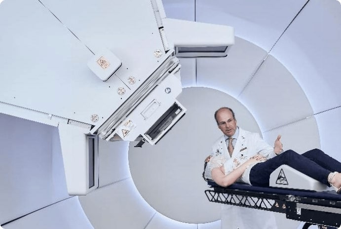
Powered by Thyrocare, Nueclear Healthcare Limited was founded in 2011. We focus on cancer diagnosis by testing, analysing, establishing and setting up diagnostic centres in the field of nuclear scanning and radiology imaging techniques. We offer a range of comprehensive solutions and assessments for accurate diagnosis of a broad spectrum of ailments.

Our Competencies

Till date, we have processed 200,000+ PET-CT scans across India. We have a vast network of over 20K+ doctors and 3K+ oncologists who trust and recommend Nueclear. Our imaging centres are led by physicians skilled in nuclear medicine to provide excellent patient care.

for-doctor-our-competencies-nuclear

Indications for PET CT Scan.

Oncology

  • In biopsy proven (Known cancer patients)
    • Staging/ metastatic workup
    • Restaging/ Response evaluation
    • Surveillance
    • To look for Recurrence
    • Post RT changes vs Viable Residual disease
  • Workup of Unknown Primary (MUO)
  • Metabolic biopsy (PET CT guided biopsy)
  • Paraneoplastic syndrome workup
  • Workup for a pathological fracture

Non-Oncological Indications

  • Cardiology- cardiac PET studies
  • Neurology- Dementia, Parkinsonism plus syndrome, intractable Epilepsy (epileptic focus localization)
  • Pyrexia of Unknown Origin (PUO) workup
  • Prosthesis infection evaluation
  • Active vs dormant/treated Granulomatous disease (TB, sarcoidosis)

Suspicion of Malignancy

  • Masses / SOLs in different organs on clinical examination or other investigations (eg. Lung mass on X-ray, breast lump on mammogram, mass in abdomen-pelvic organs on USG, etc)
  • Regional / Generalised lymphadenopathy / organomegaly
  • Suspected myeloma
  • Staging workup of patients with several cancers
  • Before surgery
  • Before Chemotherapy
  • Before radiotherapy
  • Monitoring response to treatment
  • After Surgery
  • After or during Chemotherapy
  • After or during radiotherapy
  • Follow up of treated patients with several cancers

Frequently Asked Questions

Nuclear Medicine is a clinical specialty where radiopharmaceuticals are administered to the patients for various diagnostic and therapeutic applications. Radiopharmaceuticals are radioactive drugs that consist of two components, radioactive and non-radioactive. The non-radioactive component determines the mode and organ of localization (specificity of localization in the organ of interest) and the kinetics of its biodistribution. The radioactive isotope is tagged to the non-radioactive component and the radiations emitted are used to image its invivo distribution.

Nuclear Medicine is broadly classified into “Diagnostic Nuclear Medicine” and “Therapeutic Nuclear Medicine”.

Diagnostic Nuclear Medicine involves the administration of trace quantities of radiopharmaceuticals to diagnose functional abnormalities in body tissues. It involves invivo imaging, invivo non-imaging (e.g. thyroid uptake studies, gfr estimation by plasma sampling) and in-vitro laboratory procedures (e.g. Radioimmunoassays).

The invivo imaging is broadly divided into Planar Imaging, Single Photon Emission Computed Tomography (SPECT) and Positron Emission Tomography (PET).

The most commonly used radioisotope for Planar and SPECT imaging is Technetium-99m (99mTc), having a physical half-life of 6 hours. The 99Mo-99mTc radionuclide generators are the typical routine in-house laboratory source of 99mTc availability in a nuclear medicine department. These generators have a useful life of around 7-15 days and need to be periodically replaced.

The most commonly used radioisotope for PET imaging is Fluorine-18 (18F). The 18F labelled radiopharmaceuticals are produced in a Medical Cyclotron facility and are supplied to PET centres. The short half-life of 110 minutes often does not permit its transportation to distant PET facilities.

The radiopharmaceuticals in nuclear medicine are available in a variety of forms viz. solution, colloidal solution, capsules or aerosols. Depending on its form, they are either administered intravenously, intracavitary, orally or through inhalation.

The gamma radiations emitted by the radiopharmaceuticals are detected by specialised detectors (scintillation detectors) which convert the incident radiation into light energy. This light energy is converted into electric signals and is processed further using sophisticated reconstruction algorithms to generate an image of the biodistribution of radiopharmaceutical.

Therapeutic nuclear medicine involves the administration of radiopharmaceuticals for curative (e.g. 131I-Sodium Iodide for treatment of hyperfunctioning thyroid gland in Graves’ disease, differentiated thyroid cancer) or palliative (e.g. 153mSm-EDTMP for bone pain palliation) applications.

Nuclear Medicine differs from Radiology in the following ways:

  1. Nuclear Medicine involves the internal administration of radiopharmaceuticals to the patients (radiation source is internal) while Radiology involves the use of an external source of radiation.
  2. In Nuclear Medicine, radiations emitted from the radiopharmaceutical inside the body are used for imaging. In Radiological investigations like X-ray and Computed Tomography (CT) scans, radiation produced from an X-ray generating device is directed towards the patient’s body, and radiation transmitted through the body is used for obtaining an image. The difference in the absorption of X-rays by various tissues is the basis of imaging in these investigations. Ultrasonography (USG) and Magnetic Resonance Imaging (MRI) do not involve the use of radiation.
  3. Nuclear Medicine is unique as it provides functional information of an organ or tissues in the body unlike structural information obtained from radiological investigations like X-rays, CT scans, and conventional MRI scans. Since the biochemical/functional changes precede morphological changes in the evolution of disease process, Nuclear Medicine imaging modalities can provide information on metabolic changes at the cellular level and are capable of detecting diseases much earlier than CT and MRI scans.

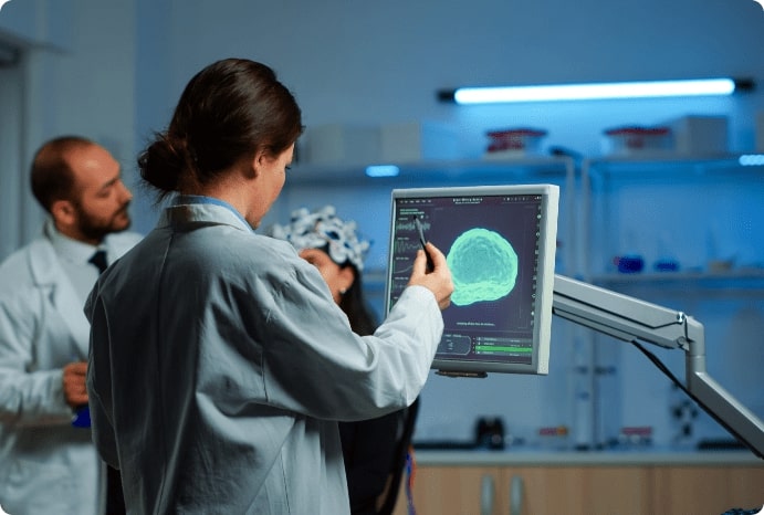
Molecular imaging is a discipline that enables the visualisation, characterization, and quantitation of biological processes taking place at the cellular level in living organisms without perturbing them. The Nuclear Medicine procedures like SPECT and PET imaging, functional MRI and optical imaging are examples of molecular imaging.

PET scan is a functional diagnostic imaging modality that involves the administration of a positron radiation-emitting radiopharmaceutical to map the various invivo biological processes. It provides clinicians with 3-dimensional images and information about how organs/tissues inside the body are functioning at the cellular and molecular levels.

When a CT scan is performed along with a PET scan, as a part of the same diagnostic workup, it is termed PET-CT fusion imaging. It uniquely combines the functional information provided by the PET scan with the anatomical information obtained from the CT scan. Fusion imaging with a CT scan thus helps localise the functional abnormality and characterise the lesion. This increases the sensitivity, specificity and overall diagnostic accuracy when compared to PET and CT alone.

PET-CT has emerged as an important complementary modality that is advancing our understanding of the underlying cause of disease and improving disease detection and management.

It provides information that may not be possible to obtain from other imaging techniques or possibly would require the use of more invasive procedures such as biopsy or surgery.

PET-CT fusion imaging helps

  • diagnose diseases in early stages, often symptoms appear, especially when other diagnostic tests are likely to give negative results.
  • assess the extent and severity of disease.
  • individualise treatment based on the unique biologic properties of the disease.
  • evaluate the effectiveness of a treatment regimen.
  • modify treatment plans in response to the altered biological behaviour of the tissue.
  • assess disease progression.
  • identify recurrence of disease and help manage ongoing care.

‘Positron radiation emitting’ radioisotope-based radiopharmaceuticals are used for PET imaging. Positrons per se are not useful for imaging as they will be absorbed within the body. However, a positron travels a short distance before losing its kinetic energy. It then annihilates with an electron to emit 2 photons of 511 keV each, which travel in opposite directions. These annihilation photons (and not positrons) are detected by the PET scanner and the light signals generated are processed by computers to provide 3-dimensional images of the tracer distribution in the body.

The most widely available PET tracers in India are 18F-Fluorodeoxyglucose (18F-FDG) & 18F-Sodium Fluoride (18F-NaF).

18F-FDG: Almost 90% of PET-CT studies are performed using 18F-FDG. Also called as the “Molecule of the Century,” 18F-FDG is a glucose analog in which the hydroxyl group at C-2 position is substituted by 18F. It is taken up by the cells via Glucose Transporter (GLUT) receptors and is subsequently phosphorylated by the Hexokinase enzyme to Fluorodeoxyglucose-6-phosphate. However, it cannot be further metabolised to deoxy fructose-6-phosphate as this step involves rearranging the carbonyl group from C-1 to C-2 position in the ring and thus getting trapped in the cells. This “metabolic trapping” of 18F-FDG forms the basis of the 18F-FDG PET-CT scan.

18F-NaF: It is used for imaging the skeletal system. It is localised in bones by binding to the hydroxyl group of the Hydroxyapatite. It has higher sensitivity and diagnostic accuracy than the conventionally used 99mTc-MDP Bone scan.

Yes! A PET Scan is an O.P.D. procedure.

18F-FDG PET-CT scan has proven efficacy in various oncological and non-oncological applications. They are summarised below:

(a) Oncological Applications

There are subtle biochemical differences between normal cells and malignant cells. In malignant cells, there is upregulation of GLUT receptors, overexpression of Hexokinase, and absence or very low levels of Glucose-6-Phosphatase. All these factors result in increased FDG uptake by tumour cells relative to the normal healthy cells. This increased FDG accumulation by malignant cells forms the basis of 18F-FDG PET-CT scan for oncological applications.

Some of the malignancies in which PET-CT modality is useful are described below:

  1. Lymphoma:
    • Routine pretreatment staging of patients with Hodgkin’s disease and Non-Hodgkin’s Lymphoma
    • Routine restaging after completing chemotherapy and radiotherapy
    • Interim assessment of treatment response for prognostication
    • Prognostication prior to bone marrow transplant/Autologous stem cell therapy
  2. Carcinoma Lung:
    • Solitary Pulmonary Nodule (SPN): To characterise a solitary pulmonary nodule > 1cm in an individual with an intermediate risk for Carcinoma lung
    • Staging of Non-Small Cell Lung carcinoma (NSCLC)
    • Treatment response assessment post-chemotherapy and radiation therapy in NSCLC
    • To assess the completeness of Radiofrequency Ablation (RFA) of carcinoma lung or pulmonary metastasis
    • Restaging of NSCLC
    • Delineation of gross tumour volume in patients scheduled for radiation therapy
  3. Head and Neck Cancer:
    • Detection of occult primary tumours in patients presenting with metastatic disease
    • Initial staging, including detection of cervical lymph node metastases when the neck nodes are not palpable and detection of distant metastases in patients with locally advanced disease
    • Detection of residual or recurrent disease
  4. Carcinoma Esophagus
    • Staging of stage I-III cancer
    • Restaging after neoadjuvant chemoradiation therapy
  5. Colorectal cancer:
    • ​​​​​​​Preoperative evaluation of patients with potentially resectable hepatic or other metastases
    • Determining the location of tumours if rising CEA level suggests recurrence
  6. Brain:
    • ​​​​​​​​​​​​​​To identify anaplastic transformation of non-enhancing low-grade gliomas
    • To grade gliomas non-invasively and guide biopsy
    • To differentiate radiation necrosis from disease recurrence
  7. Thyroid cancer:
    • ​​​​​​​​​​​​​​Detection of residual or recurrent differentiated thyroid cancer when serum thyroglobulin is elevated and radioiodine scan is negative
    • Staging of poorly differentiated/anaplastic thyroid carcinoma
  8. Carcinoma Breast
  9. Genitourinary cancer:
    • ​​​​​​​​​​​​​​Initial treatment planning, including determination of nodal status and systemic spread.
    • Detection of residual or recurrent disease
  10. Musculoskeletal system:
    • ​​​​​​​​​​​​​​Staging and interim response evaluation of PNET / Ewing's Sarcoma
    • To assess treatment response to therapy when pre-treatment PET-CT shows FDG avid lesions
    • To assess sarcomatous change in osteochondroma, grade it non-invasively, and direct biopsy
    • To assess the completeness of RFA in cases of Osteoid osteoma
    • To differentiate plasmacytoma from multiple myeloma
  11. Neuroblastoma:
    • ​​​​​​​​​​​​​​To stage the disease
    • To assess treatment response
    • To restage the disease
      1. Non-Oncological Applications: Increased glycolysis in the inflammatory cells forms the basis of 18F-FDG PET-CT imaging for various non-oncological pathologies such as aseptic inflammatory processes as well as in a wide variety of infections. The non-oncological applications of FDG PET-CT scan are as follows:
        • Pyrexia of Unknown Origin (PUO):
          Three categories that account for the majority of PUO are infections, malignancies, and collagen-vascular or autoimmune diseases. Early identification and localization of an infectious or inflammatory process can be critical for the management of these patients. Because of its high sensitivity in detecting malignant lesions, infections like Tuberculosis, as well as various inflammatory processes, FDG-PET has the potential to play a central role in the management of patients with PUO.
        • Epilepsy:
          In cases of temporal lobe epilepsy, increased tracer uptake is noted in the 99mTc-ECD ictal study. The 18F-FDG interictal scan is performed to increase diagnostic specificity.
        • Dementia:
          In its early stages, the identification and differential diagnosis of dementia is especially challenging, because of the difficulty in distinguishing it from the mild cognitive decline associated with normal ageing. The specific patterns of altered metabolism are suggestive of dementia. FDG PET may be the ideal test for selecting the appropriate patients for treatment when the disease process is at the molecular level and before structural alterations have taken place.
        • Sarcoidosis:
          To evaluate the extent of disease in diagnosed cases of sarcoidosis and assessing treatment response
        • Vasculitis involving major vessels:
          To evaluate the extent of disease and monitor the effectiveness of therapy
        • Cardiology:
          To assess viability in dysfunctional myocardial segments

Recently, 18F-NaF PET-CT scan has gained popularity as an alternative to the conventionally used 99mTc-MDP Bone scans due to the following advantages:

  • 18F-NaF PET-CT has higher sensitivity and specificity in distinguishing benign from malignant lesions as compared to MDP Bone scan.
  • It provides sharper images with higher resolution than conventional planar bone scan and SPECT
  • The Fluoride bone scan requires 90 minutes for its completion while the MDP bone scan requires 3-4 hours.
  • Low dose CT associated with PET-CT study increases its specificity and thus obviates the need of an additional diagnostic CT or MRI scan. The indications of the NaF bone scan are as follows:
    • Evaluation of skeletal metastases in a case of carcinoma prostate
    • Evaluation for skeletal metastases in stage III and IV carcinoma breast and symptomatic cases of Stage I and II
    • Evaluation of cases of osteogenic sarcoma
    • Assessment of treatment response in metabolic bone diseases
    • Evaluate a case of osteoid osteoma prior to and post RFA for assessing completion of ablation
    • Evaluation of low backache
    • Prior to radioisotopic bone pain palliation therapy

  • The patient should strictly fast for at least 6 hours. An increase in the blood sugar levels after a meal may competitively inhibit the uptake of FDG (a glucose analog) in the lesions and thus decrease the diagnostic sensitivity and overall accuracy of the modality. Also, it causes the endogenous release of insulin which results in increased tracer uptake in skeletal muscles. This altered biodistribution of FDG makes the study suboptimal for qualitative and quantitative assessment. The patient may, however, be permitted to drink plain water.
  • The patient should not undertake any intense and strenuous physical activity or exercise for 24 hours before the scan as it results in increased uptake in the skeletal muscles (i.e. altered biodistribution).
  • The patient should carry all the relevant medical records, reports, film, and/or CD of X-rays, CT, MRI, PET-CT or any other investigation done to date on the day of the study.
  • An adult attendant should accompany the patient at the time of the scan. The patient should not be accompanied by children and pregnant women.
  • The patient should wear loose and comfortable clothing on the day of the scan. The patient should not wear any metallic objects, jewellery or valuables.
  • For nursing and pregnant patients:
    • If a patient is nursing, pregnant or thinks that she may be pregnant, she should inform the staff at the time of appointment.
  • For diabetic patients only (in addition to the earlier instructions):
    • The patient should not take any anti-diabetic medicines (tablets/insulin injections) on the day of the scan.
    • The patient should have good glycemic control. A fasting blood sugar of <150 mg/dl is desirable.

  • The patient need not be fasting at the time of the study.
  • Diabetic patients can have breakfast and their anti-diabetic medicines. High blood sugar does not interfere with the results of the scan.
  • Rest of the instructions to be followed are the same as those of the 18F-FDG PET-CT scan (mentioned in the previous section).

  • The patient is injected with the radiopharmaceutical and asked to stay in a separate room for approximately 60-90 minutes. This mandatory waiting period is required for the radiotracer to localise in the target tissues. The patients are advised to restrict physical movements and avoid talking to others while present in the room. During this resting period, unless recommended by the staff, no attendants or relatives would be permitted to stay with the patients.
  • The patient may be given oral contrast to drink. This will help to obtain more informative images of the abdomen. Just before starting the scan, the patient would be asked to pass urine.
  • The patient is then subjected to scanning and during the scanning is expected to remain still for 15-20 minutes.
  • After the completion of scanning, the PET-CT scan is reviewed for the quality and adequacy of the study. It may be repeated if required.
  • The PET-CT scan is finally examined by an experienced Nuclear Medicine Physician and findings are reported.

Yes! The PET-CT procedures are safe, painless, non-invasive, and cost-effective.

PET-CT scan procedures are rarely associated with any significant discomfort or side effects.

done in tandem. For a PET scan, the radiopharmaceutical is injected in small (tracer) quantities. It is excreted from the body through urine. The un-excreted radiopharmaceutical decays with a short half-life (110 minutes). Thus, the amount of radiation exposure received by the patient is very low.

Sr No Procedure Effective Dose (mSv)
1 18F FDG PET scan 7
2 99mTc Bone Scan 4.4
3 Whole-body CT scan 8-30
4 CT head 2
5 CT chest 8
6 CT abdomen 10
7 CT pelvis 10
8 Coronary Angiography 5-15
9 Mammography 0.13
10 X-Ray chest 0.04
11 X-Ray Abdomen 0.7

Source: http://hps.org/hpspublications/articles/dosesfrommedicalradiation.html

The estimated effective dose from a typical PET scan is 8 milliSievert (mSv). It is equivalent to the radiation dose received from the natural environment in 3 years. The effective dose from CT has a very wide range (8-30 mSv) depending on the type of the test, the region of the body scanned, and the purpose of the test.

The radiopharmaceuticals used for PET scans are absolutely safe and have no reported allergic reactions.

The CT scan done as a part of the PET-CT procedure may be performed with or without contrast enhancement. These contrast agents are known to cause allergic reactions in few patients as seen with any other contrast-enhanced CT procedure. The routinely used non-ionic contrast media are safe. However, in very few cases some side effects may be noticed as follows:

Minor Reactions: Itching, rashes, chills, nausea and vomiting. They are self-limiting and require no treatment.

Moderate reactions: They include dyspnea, tachycardia, generalised erythema and mild hypotension. The chance of such reactions is 1 in 1,000 i.e 0.1%.

Severe Reactions: They occur rarely and include convulsion, cardiopulmonary arrest, profound hypotension and arrhythmias. One in 1,00,000 studies (i.e. 0.01%) may lead to death.

Yes! However, it is recommended that the patient does not breastfeed her baby for 6-8 hours after the scan has been performed as small amounts of the administered radiopharmaceutical might be excreted in breast milk. It is advisable to collect expressed breast milk before the radiotracer injection so that it can be used to feed the baby.

Generally, there are no restrictions on patients’ social behaviour after a PET-CT scan. The patients may resume their routine activities immediately after the scan is over. However, it is advisable to avoid prolonged contact with infants, children and pregnant women for at least 6 hrs after the scan.

The PET-CT scan results are usually available within 2 days.

The accessibility of PET-CT scans has led to the widespread availability of metabolic scans which have reliable and better diagnostic results.

With the development of new specific radiotracers and targeted therapies, improvement in the resolution of imaging systems and fusion imaging with MRI, the existing list of applications of PET scan in clinical practice is likely to increase.

We are providing you this sweet experience by using cookies

Accept Privacy Policy